

Anti Smooth Muscle Antibody (ASMA) Test: Clinical Meaning, Titer Ranges, and Diagnostic Role
Introduction
When routine blood work reveals elevated liver enzymes (such as ALT and AST), physicians often launch a diagnostic investigation to determine the root cause of hepatic inflammation. Among the most informative specialized blood tests ordered during this evaluation is the Anti Smooth Muscle Antibody (ASMA) test.
Understanding what an anti-smooth muscle antibody result signifies can feel overwhelming, especially when terms like “autoimmunity,” “titers,” and “hepatitis” enter the conversation. This comprehensive guide breaks down the science behind ASMA, its primary role in identifying conditions like Autoimmune Hepatitis (AIH), how to interpret reference ranges, and what steps follow a positive test.
What Is an Anti Smooth Muscle Antibody?

An Anti Smooth Muscle Antibody (ASMA)—sometimes referred to simply as a Smooth Muscle Antibody (SMA)—is a type of autoantibody. Under normal circumstances, the human immune system produces protective proteins called antibodies to detect and destroy foreign pathogens such as viruses, bacteria, and parasites.
In individuals with an autoimmune predisposition, the immune system loses its ability to distinguish between “self” and “non-self”. It mistakenly creates autoantibodies that target the body’s own healthy cellular structures.
The Role of Actin and Cytoskeletal Proteins
Anti-smooth muscle antibodies target components within the cell’s structural framework, known as the cytoskeleton. Specifically, ASMAs direct their attack against microfilaments, primarily a protein called F-actin (filamentous actin).
Because smooth muscle tissue is abundant in blood vessels and organs throughout the body—including the blood vessels surrounding and inside the liver—these autoantibodies serve as critical biological markers for immune-mediated tissue damage, particularly in liver parenchymal tissue Strongest Muscle in the Human Body.
Why Is the ASMA Test Performed?
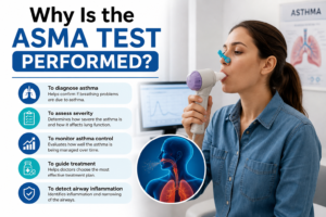
Healthcare providers do not typically include the ASMA test in basic wellness screenings. Instead, it is requested as part of a targeted autoimmune liver panel when a patient exhibits unexplained symptoms or abnormal liver biochemistry.
Unexplained Liver Symptoms / Elevated ALT & AST
                      │
                      ▼
         Initial Diagnostic Panel
  (Viral Hepatitis, Metabolic & Alcohol Screening)
                      │
                      ▼
       If Negative / High Suspicion
                      │
                      ▼
            Autoimmune Liver Panel
          (ASMA + ANA + IgG Levels)
Primary Diagnostic Indications
-
Investigating Unexplained Liver Inflammation: If routine blood tests reveal chronically elevated alanine aminotransferase (ALT) or aspartate aminotransferase (AST), doctors must rule out viral infections (like Hepatitis B or C), non-alcoholic fatty liver disease, and drug-induced liver injury.
-
Diagnosing Autoimmune Hepatitis (AIH): ASMA is one of the classic hallmark markers used to identify Type 1 Autoimmune Hepatitis.
-
Differentiating Liver Diseases: Symptoms of liver dysfunction often overlap across various conditions. ASMA helps differentiate AIH from other biliary or cholestatic disorders such as Primary Biliary Cholangitis (PBC) and Primary Sclerosing Cholangitis (PSC).
-
Monitoring Treatment Response: In patients with established AIH, tracking antibody titers over time alongside liver enzyme trends provides valuable insight into whether immunosuppressive therapy is successfully dampening the autoimmune response.
Symptoms That Prompt an ASMA Test
Because early-stage liver inflammation can be subtle, symptoms may develop slowly over several months or emerge suddenly. Doctors routinely evaluate for anti-smooth muscle antibodies when patients exhibit a combination of the following clinical signs:
-
Persistent Fatigue: Severe, unrelenting exhaustion that does not improve with rest—often the earliest and most common symptom of autoimmune liver disease.
-
Jaundice: Yellowing of the skin and whites of the eyes (sclera) caused by bilirubin accumulation in the blood.
-
Abdominal Discomfort: Dull aching or fullness in the upper right quadrant of the abdomen, where the liver resides.
-
Joint and Muscle Pain (Arthralgia): Systemic aches in multiple joints, characteristic of generalized autoimmune activity.
-
Pruritus (Itchy Skin): Generalized itching without a visible skin rash, often linked to impaired bile clearance.
-
Nausea and Appetite Loss: Unexplained digestive distress, sometimes accompanied by unintended weight loss.
-
Dark Urine and Pale Stools: Changes in excretion colors indicative of biliary tree or hepatocyte stress.
Anti-Smooth Muscle Antibodies and Autoimmune Hepatitis (AIH)

To fully appreciate the clinical utility of the ASMA test, it is essential to understand Autoimmune Hepatitis (AIH). AIH is a chronic, non-contagious liver disease in which T-lymphocytes and autoantibodies attack hepatocytes (liver cells), causing continuous inflammation, necrosis, and potentially progressive fibrosis or cirrhosis if untreated.
Autoimmune Response against Hepatocytes
                  │
                  ▼
         Chronic Inflammation
                  │
                  ▼
         Fibrosis (Tissue Scarring)
                  │
                  ▼
  Cirrhosis / End-Stage Liver Disease (If untreated)
Autoimmune hepatitis is broadly classified into two distinct subtypes based on specific circulating autoantibody profiles:
Type 1 Autoimmune Hepatitis
-
Prevalence: Accounts for roughly 80% to 90% of all AIH cases worldwide.
-
Target Demographics: Can occur at any age but is most frequently diagnosed in late adolescence to early adulthood, as well as in mid-life, with a strong female predominance (~80% of cases).
-
Key Serological Markers: Marked by the presence of Anti-Smooth Muscle Antibodies (ASMA), Antinuclear Antibodies (ANA), or both. Elevated serum Immunoglobulin G (IgG) levels are also typical.
Type 2 Autoimmune Hepatitis
-
Prevalence: Less common, accounting for 10% to 20% of AIH cases.
-
Target Demographics: Predominantly affects children, infants, and young adolescents.
-
Key Serological Markers: Characterized by Anti-Liver Kidney Microsomal Type 1 (anti-LKM1) antibodies or Anti-Liver Cytosol Type 1 (anti-LC1) antibodies, with ASMA typically remaining negative.
How the ASMA Test Is Performed and Calculated
The ASMA blood test requires a standard venipuncture. No fasting or complex preparation is strictly required, though patients should inform their healthcare provider about all medications, supplements, and recent viral illnesses.
Blood Sample Collection (Venipuncture)
                  │
                  ▼
       Laboratory Analysis (IFA)
                  │
                  ▼
      Serial Dilution Procedure
   (1:20 ──► 1:40 ──► 1:80 ──► 1:160)
                  │
                  ▼
 Final Titer Determination (Highest Positive Ratio)
Laboratory Methodology: Indirect Immunofluorescence Assay (IFA)
The gold-standard laboratory technique for detecting ASMA is the Indirect Immunofluorescence Assay (IFA).
-
The patient’s blood serum is applied to a tissue slide containing smooth muscle targets (often rodent stomach, kidney, or liver sections).
-
If smooth muscle autoantibodies are present in the serum, they bind to the smooth muscle structures on the slide.
-
A fluorescent dye-tagged antibody is added, which adheres to the human autoantibodies.
-
Pathologists view the slide under a fluorescence microscope to observe characteristic staining patterns (such as vascular, visceral, or glomerular patterns).
Understanding Titer Ratios
Laboratory results are often reported as a titer ratio. A titer represents how many times a patient’s serum can be diluted with saline while still showing detectable autoantibody fluorescence under the microscope.
-
Dilutions progress exponentially: 1:20, 1:40, 1:80, 1:160, 1:320, and higher.
-
A higher denominator indicates a greater concentration of autoantibodies in the bloodstream. For instance, a titer of 1:160 means antibodies are still detectable even when diluted 160 times, signifying a stronger antibody response than a 1:20 dilution.
Interpreting ASMA Test Results
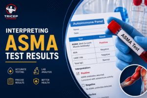
Because reference ranges and laboratory units can vary slightly depending on whether a lab uses indirect immunofluorescence (titers) or solid-phase immunoassay ELISA units (measured in Units/mL), test results should always be interpreted using the specific laboratory’s reference interval.
| Result Classification | Immunofluorescence Titer (IFA) | ELISA Signal (Units/mL) | Clinical Interpretation |
| Negative | < 1:20 |
< 20 U/mL |
Normal baseline. Autoantibodies are not detected in significant quantities. |
| Weak / Borderline | 1:20 to 1:40 |
20 to 30 U/mL |
Low autoantibody concentration. May reflect early AIH, transient viral response, or non-specific inflammation. |
| Moderate to High Positive | ≥ 1:80 |
> 30 U/mL |
Clinically significant autoantibody elevation. Strongly supports Autoimmune Hepatitis Type 1 when combined with elevated liver enzymes. |
What Does a Negative Result Mean?
A negative ASMA result (< 1:20) generally suggests that smooth muscle autoantibodies are absent or below detectable levels. While this significantly lowers the probability of Type 1 Autoimmune Hepatitis, it does not rule out liver disease entirely. Some individuals may have seronegative AIH, Type 2 AIH, or non-autoimmune liver disorders.
What Does a Borderline/Weak Positive Result Mean?
A low-titer result (1:20 or 1:40) is common and must be interpreted cautiously. Low-level autoantibodies can briefly appear due to recent viral infections, systemic immune activation, or minor tissue inflammation. Re-testing in 8 to 12 weeks is often recommended to see if levels rise or return to baseline.
What Does a High Positive Result Mean?
A titer of 1:80 or higher is considered strongly positive and clinically meaningful. When paired with elevated ALT/AST and high total IgG levels, a strong ASMA positive significantly raises the clinical suspicion for Autoimmune Hepatitis.
Causes of a Positive ASMA Test Beyond Autoimmune Hepatitis
While ASMA is most closely associated with Type 1 AIH, a positive test is not 100% specific to a single condition. Doctors evaluate the broader clinical picture because smooth muscle antibodies can appear in several other medical contexts:
                     Causes of Positive ASMA
                                │
  ┌─────────────────────────────┼─────────────────────────────┐
  ▼                             ▼                             ▼
Autoimmune Liver Diseases   Viral Infections           Other Conditions
• Autoimmune Hepatitis (AIH) • Hepatitis B & C          • Systemic Lupus (SLE)
• Primary Biliary            • EBV (Mononucleosis)     • Drug-Induced Injury
  Cholangitis (PBC)          • Cytomegalovirus (CMV)   • Cancers (rare)
-
Other Autoimmune Liver Conditions:
-
Primary Biliary Cholangitis (PBC): Roughly 10% to 22% of PBC patients may test positive for ASMA alongside Antimitochondrial Antibodies (AMA).
-
Primary Sclerosing Cholangitis (PSC): Low-to-moderate ASMA titers can occasionally occur in PSC patients, particularly in overlap syndromes.
-
-
Viral Infections:
-
Chronic Hepatitis B or C: Viral injury to liver cells can release intracellular actin into circulation, triggering secondary antibody production.
-
Epstein-Barr Virus (EBV) & Cytomegalovirus (CMV): Transient ASMA positivity can occur during acute infectious mononucleosis and generally resolves once the viral infection clears.
-
-
Non-Hepatic Autoimmune Disorders:
-
Conditions such as Systemic Lupus Erythematosus (SLE), rheumatoid arthritis, or autoimmune thyroiditis can sometimes cause low-level cross-reactive autoantibodies.
-
-
Drug-Induced Liver Injury (DILI):
-
Certain prescription medications (such as minocycline, nitrofurantoin, or infliximab) can trigger a drug-induced autoimmune-like hepatitis complete with positive ASMA titers that subside after stopping the drug.
-
Diagnostic Workup: The Complete Autoimmune Liver Profile
Because an ASMA test alone cannot confirm a diagnosis, hepatologists and gastroenterologists utilize a comprehensive multi-step evaluation.
Step 1: Serological Testing (ALT, AST, ASMA, ANA, IgG)
                  │
                  ▼
Step 2: Non-Invasive Imaging (Ultrasound / FibroScan)
                  │
                  ▼
Step 3: Liver Biopsy (Histological Evaluation)
                  │
                  ▼
Step 4: IAIHG Scoring & Diagnostic Confirmation
1. Comprehensive Blood Panels
-
Liver Function Tests (LFTs): Evaluates ALT, AST, Alkaline Phosphatase (ALP), Total Bilirubin, and Albumin.
-
Complementary Autoantibodies: Checks Antinuclear Antibody (ANA), Anti-LKM1, Antimitochondrial Antibody (AMA), and Anti-Soluble Liver Antigen (Anti-SLA).
-
Anti-F-Actin ELISA: A refined assay specifically targeted at filamentous actin, offering higher diagnostic specificity for AIH than broader ASMA immunofluorescence.
-
Serum Immunoglobulin G (IgG): High levels of IgG reflect hypergammaglobulinemia, a classic feature of active AIH.
2. Diagnostic Imaging
Abdominal ultrasound, CT scans, or Magnetic Resonance Cholangiopancreatography (MRCP) are utilized to assess liver architecture and exclude gallstones, bile duct obstructions, or fatty liver disease.
3. Liver Biopsy: The Gold Standard
If blood markers and symptoms suggest Autoimmune Hepatitis, a liver biopsy is typically recommended to confirm the diagnosis. A pathologist examines a small tissue sample under a microscope looking for hallmark features:
-
Interface Hepatitis: Inflammation spilling from the portal tracts into the surrounding liver parenchyma.
-
Lymphoplasmacytic Infiltrates: An abundance of plasma cells invading the tissue.
-
Hepatocyte Rosettes and Fibrosis: Structural changes indicating cellular injury and regenerative attempts.
Treatment and Management of Autoimmune Hepatitis
When a positive ASMA result leads to a confirmed diagnosis of Autoimmune Hepatitis, early medical treatment is key. The primary goal of therapy is to induce and maintain complete clinical and biochemical remission, preventing progressive scarring.
Initial Remission Induction (Corticosteroids e.g., Prednisone)
                  │
                  ▼
Long-Term Maintenance (Steroid-Sparing Agents e.g., Azathioprine)
                  │
                  ▼
Ongoing Monitoring (Periodic LFTs, IgG, and ASMA Titers)
-
Induction Phase: High-dose systemic corticosteroids (such as Prednisone or Prednisolone) or liver-targeted steroids (like Budesonide) are initiated to suppress active liver inflammation quickly.
-
Maintenance Phase: Once liver enzymes begin normalizing, steroid-sparing immunosuppressive medications—most commonly Azathioprine—are introduced to maintain long-term remission while minimizing steroid side effects.
-
Monitoring & Prognosis: Remission is monitored by checking liver enzymes (ALT/AST) and IgG levels, alongside periodic ASMA titers. With early intervention and consistent medication adherence, the long-term outlook for individuals with Autoimmune Hepatitis is overwhelmingly positive, allowing most patients to lead full, healthy lives.
Frequently Asked Questions (FAQs)
1. Is an Anti Smooth Muscle Antibody test the same as an Anti-F-Actin test?
They are closely related, but not identical. ASMA tests look for antibodies against a broad group of smooth muscle cytoskeletal proteins using immunofluorescence. The anti-F-actin test specifically isolates and measures antibodies against filamentous actin (the primary target in AIH) using ELISA technology, offering higher diagnostic specificity.
2. Can a positive ASMA result go back to negative?
Yes. If the positive ASMA result was triggered by a temporary viral infection (such as EBV or acute hepatitis), antibody levels usually decline and disappear once the infection clears. In patients with Autoimmune Hepatitis, effective immunosuppressive treatment often reduces antibody titers significantly, sometimes bringing them below detectable lab limits.
3. Does a positive ASMA test mean I have liver cirrhosis?
No. A positive ASMA test simply indicates the presence of autoantibodies in your blood; it does not directly measure liver damage or stage fibrosis. Determining whether cirrhosis or scarring is present requires imaging tests (such as FibroScan or elastography) or a liver biopsy.
4. What is considered a normal ASMA titer?
A normal ASMA result is negative, which is typically reported as a titer of less than 1:20 (or < 20 U/mL on an ELISA scale). Values at or below this threshold mean autoantibodies were not detected in significant amounts.
5. Can diet or lifestyle changes lower ASMA titers?
There is no specific diet or natural supplement proven to lower anti-smooth muscle antibody levels directly. Because ASMA is generated by an underlying autoimmune reaction, pharmaceutical immunosuppression guided by a physician is necessary to manage antibody activity and liver inflammation. However, eating a balanced, liver-friendly diet, avoiding alcohol, and maintaining a healthy weight help reduce overall stress on the liver.
6. Can you have Autoimmune Hepatitis if your ASMA test is negative?
Yes. Approximately 10% to 20% of Autoimmune Hepatitis cases are Type 2 AIH, which involves different autoantibodies (anti-LKM1 or anti-LC1) rather than ASMA. Additionally, a small percentage of individuals have seronegative AIH, where classic autoantibodies are absent despite histological evidence of autoimmune liver inflammation.